
Reviewed by clinicaobesidadenvejecimiento.com · Last review:

Diagnosing biopolymers combines three pieces: the medical history (what was injected, when and where), physical examination of the area, and imaging, especially MRI, which shows the extent of the material and the affected tissue. Lab tests help assess inflammation and general health. With that information the stage of the disease and the plan are defined: medical management, localized removal or both. Removing 100% of the material is not always possible, so an accurate diagnosis is the basis of safe treatment.
- Medical history
- MRI
- Plan by stage
Why diagnosis is the first step
Filler substances do not stay in one spot: they spread through tissue, can migrate and cause chronic inflammation. In the experience of the Hospital General de México, care always begins by locating the material and understanding the patient’s condition before any surgery. Without that map, removal can be incomplete or carry more risk.
1. Medical history
The consultation starts with specific questions that guide the diagnosis:
- Which substance was applied, if known (liquid silicone, oil, paraffin, polyacrylamide or other).
- When and where it was applied, how many sessions and who applied it.
- What symptoms there are, since when and whether they are increasing.
- Previous treatments, illnesses and current medications.
Many people do not know exactly what was injected. COFEPRIS warns that these substances are often applied in unauthorized places, so the history is always complemented with tests.
2. Physical examination
The doctor checks the injected area and nearby areas: swelling, hardening, nodules, color changes, ulcers or migration of the material. Other parts of the body are also examined, because the reaction can spread.
3. MRI
This is the key test. It shows where the material is, how much there is, how deep it goes and whether it has migrated, as well as how inflamed the neighboring tissue is. The Hospital General de México uses it as part of its multidisciplinary protocol, and the Latin American series of 250 patients uses it to classify patients by stage.
| Test | What it shows | When it is used |
|---|---|---|
| MRI | Extent, depth and migration of the material; tissue inflammation | Basis for planning treatment |
| Ultrasound | Initial assessment of nodules and fluid collections | As support or follow-up |
| Lab tests | Inflammation, kidney and liver function, general health | Before any procedure |
4. Disease stage and plan
With the history, examination and tests, the specialist defines the clinical stage. Published series propose treatment by stage (Cir Plast Iberolatinoam 2022), and there is a gluteal-specific staging system that helps plan surgery.
Medical management
- Control of inflammation
- Foundation of treatment at every stage
- Prepares the tissue for possible surgery
Localized removal
- Defined masses or areas
- When the tissue is ready
- With laser support and reconstruction if needed
What diagnosis can realistically tell you
Coiffman recommends resecting only limited areas, because removing all the material is usually not possible without damaging healthy tissue. A good diagnosis helps set expectations: the goal is to remove what is safe and control symptoms, not to promise total removal.
Evidence on the specific use of laser in these cases is limited (2025 systematic review), so it is used as a supporting tool within a complete plan.
Myth and fact
Myth: “Touching the area is enough to know how much material there is.”
Fact: Only MRI shows the real extent and whether it has migrated.
Myth: “If I do not remember what was injected, it cannot be treated.”
Fact: The history is complemented with imaging that locates the material.
Myth: “Diagnosis shows that everything can be removed.”
Fact: Coiffman recommends resecting limited areas; removing 100% is not always possible.
Myth: “Tests are unnecessary if there are already symptoms.”
Fact: They are the basis for planning safe treatment (Hospital General de México).
Want to know what you have and where it is?
Book an assessment: we review your history and the area and tell you which tests you need for a safe plan.
Book an assessmentFrequently asked questions
Which test matters most?
MRI, because it shows the extent, depth and migration of the material.
Is ultrasound useful?
It helps with initial assessment and follow-up, but MRI gives the most complete map.
What are the lab tests for?
To assess inflammation and general health before any procedure.
Is the diagnosis painful?
The consultation and imaging are non-invasive.
Can I be treated without tests?
It is not advisable; a safe plan depends on locating the material (Hospital General de México).
What happens after the diagnosis?
The stage and plan are defined: medical management, localized removal or both.
Keep reading
Downloadable quick guide. The diagnostic steps and what each test shows, in one image.
Download infographic (PNG)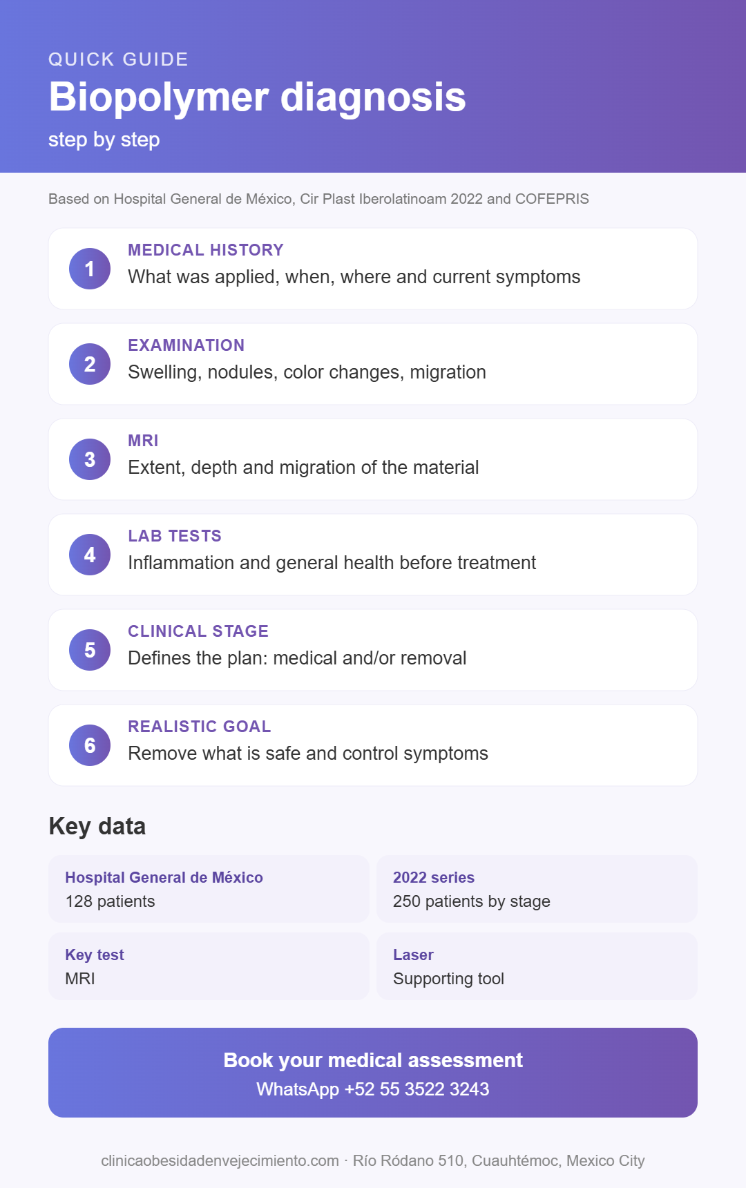
Sources
- Gordillo-Hernández J, et al. Multidisciplinary approach to filler substance disease. Hospital General de México. Cir Plast Iberolatinoam. 2013;39(3). SciELO
- Allogenosis from biomaterials, our experience. Cir Plast Iberolatinoam. 2022. SciELO
- A staging system for gluteal foreign body reaction to injectables. J Plast Reconstr Aesthet Surg. ScienceDirect
- Coiffman F. Iatrogenic allogenosis: a new disease. Cir Plast Iberolatinoam. 2008;34(1). SciELO
- COFEPRIS. Statement 96/2023 on filler substances. gob.mx
- Foreign body granulomas related to cosmetic fillers: a systematic review. 2025. PubMed Central